

Home › Trichology for beginners › Chapter 06
Trichogramand Trichoscopy
A trichogram examines plucked hairs under a microscope, while trichoscopy magnifies the scalp without removing hair. Learn what each test reveals and when it is used.
 Est. 1989 — Spain
A trichogram examines hairs removed from a selected area of the scalp under a microscope, while trichoscopy magnifies the hairs, follicular openings and scalp surface without removing them.
The two methods are complementary rather than interchangeable, and their findings must be interpreted alongside the person’s history and clinical examination.
What hair and scalp analysis means
“Hair analysis” can refer to several different procedures. In professional trichology, it normally means the visual and microscopic assessment of the hair and scalp: looking at the scalp surface, hair density, follicular openings, hair-shaft diameter, breakage and other visible patterns.
This should not be confused with commercial tests that claim to diagnose nutritional or systemic conditions by measuring minerals in a hair sample. A clinical trichogram and trichoscopy examine the physical characteristics of the hair, roots and scalp.
The essential difference: trichoscopy examines the hair and scalp in place, while a traditional trichogram examines a small sample of plucked hairs under a microscope.
Two complementary techniques
They answer different questions. One provides a magnified view of the scalp and visible hairs; the other provides microscopic information about the roots and growth phases of a sampled group of hairs.
Trichoscopy
Dermoscopy applied to hair & scalp
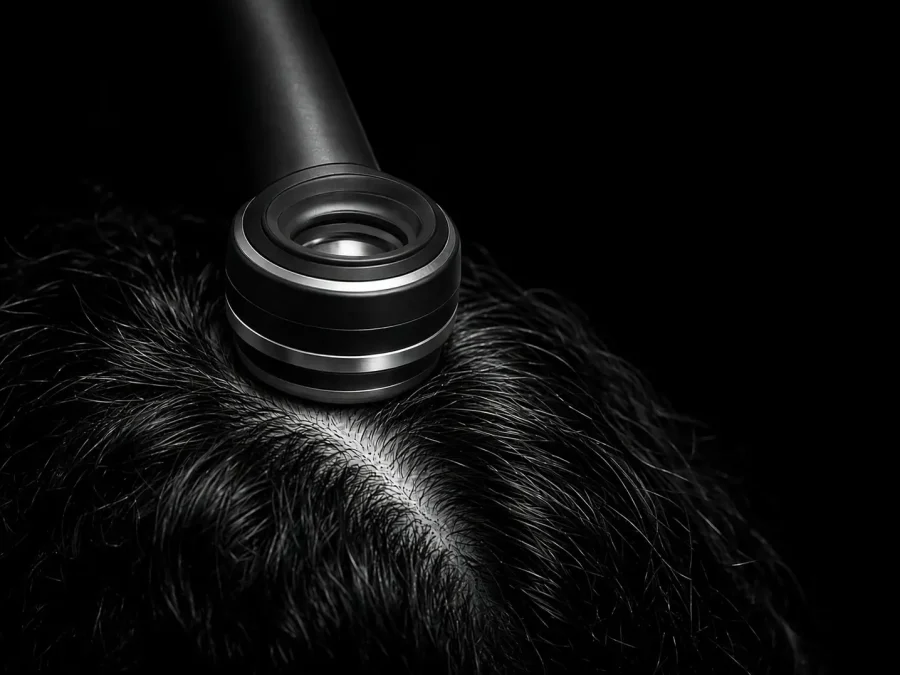
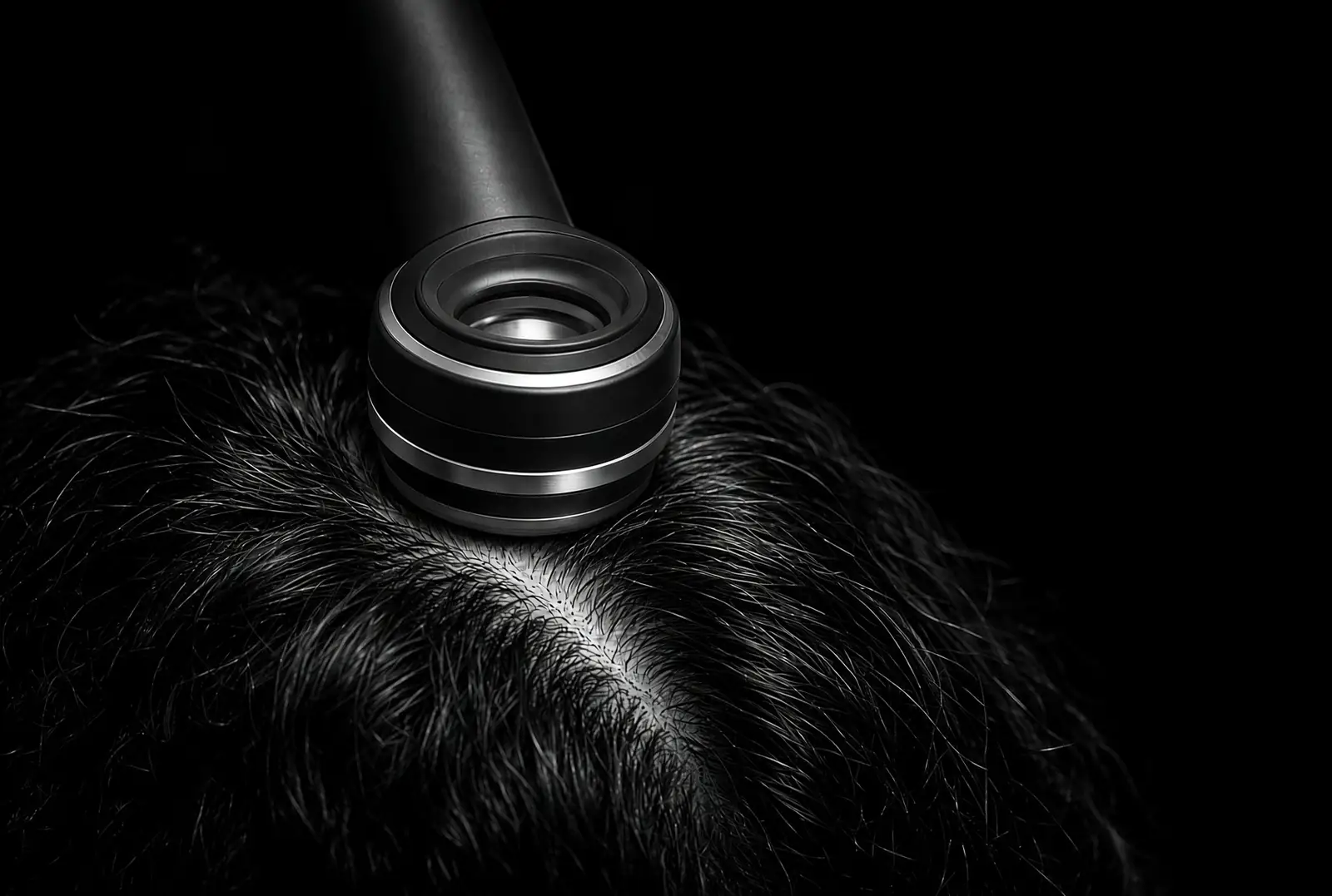
A handheld dermoscope or digital video system magnifies the scalp surface, follicular openings and emerging hair shafts. Images can be stored and compared during follow-up.
Non-invasive · no samples · repeatable
The trichogram
Microscopic examination of plucked hairs
A small sample of hairs is removed and examined under a microscope to assess the roots, shafts and distribution of hairs across different stages of the growth cycle.
Semi-invasive · microscopic · sampled hairs
What is trichoscopy?
Trichoscopy is dermoscopy applied to the hair and scalp. The instrument combines magnification with controlled lighting, allowing the examiner to see structures and patterns that are difficult or impossible to assess with the naked eye.
The examination is normally quick and painless, and no hairs need to be removed. Depending on the equipment, images may be viewed through a handheld instrument or displayed and stored digitally.
During trichoscopy, the examiner may assess:
- Variation in hair-shaft diameter and signs of miniaturisation.
- The number of hairs emerging from individual follicular units.
- Broken, tapered, coiled or otherwise altered hairs.
- Empty follicular openings and changes around the follicles.
- Scaling, redness and other changes to the scalp surface.
- Vascular and pigment patterns.
- Features that may suggest scarring or non-scarring hair loss.
These findings form a magnified pattern rather than a diagnosis by themselves. The same feature can have different meanings depending on its distribution, the person’s symptoms and the rest of the clinical examination.
Useful for follow-up: standardised trichoscopic photographs can document changes in density, hair diameter and scalp appearance over time.
What is a trichogram?
A trichogram is the microscopic examination of hairs removed from selected areas of the scalp. A small group of hairs is plucked so that the proximal end — including the root region — can be examined.
The examiner can classify the sampled hairs according to their appearance and stage of the hair cycle. Depending on the purpose of the examination, the trichogram may estimate the proportion of hairs in the anagen growth phase, the transitional catagen phase and the resting or shedding telogen phase.
A trichogram may provide information about:
- The distribution of sampled hairs across the growth cycle.
- Roots that appear altered, dystrophic or broken.
- Changes consistent with increased shedding.
- Certain abnormalities affecting the hair shaft.
- Differences between samples taken from separate scalp areas.
Its reliability depends on correct sampling, preparation, the area selected and experienced interpretation. The result describes the hairs included in that particular sample and must be considered within the wider clinical picture.
Does a trichogram hurt? Because several hairs are plucked, it can cause a brief pulling sensation or short-lived discomfort. It is less comfortable than trichoscopy but does not normally require an anaesthetic.
What happens during the examination?
- Clinical history: the specialist asks about the onset and pattern of the problem, family history, illnesses, medication, nutrition, stress, styling practices and scalp symptoms.
- Visual examination: the scalp is inspected for the distribution of thinning, inflammation, scale, broken hairs and other relevant signs.
- Trichoscopy: representative areas are examined under magnification and images may be recorded for comparison.
- Trichogram when indicated: a small sample of hairs is removed and examined microscopically.
- Interpretation: the instrumental findings are combined with the clinical history and examination.
If the cause remains uncertain, a dermatologist may recommend additional investigations such as blood tests, fungal testing or, in selected cases, a scalp biopsy.
How should you prepare?
Preparation depends on the test and the protocol used by the clinic. Unless instructed otherwise, keep your usual routine and avoid covering the scalp with concealing fibres, heavy oils or excessive styling residue on the day of the examination.
If a trichogram is planned, follow the examiner’s specific preparation and washing instructions because these can affect sampling and may vary between protocols.
What can a hair and scalp analysis reveal?
Together, these techniques can help an experienced examiner evaluate:
- Hair density: how the visible hairs are distributed across different scalp areas.
- Follicular units: whether one or several hairs emerge from individual openings.
- Hair diameter: whether some areas show finer or progressively miniaturised hairs.
- Growth-cycle activity: the estimated distribution of sampled hairs across anagen, catagen and telogen phases.
- Hair-shaft damage: fractures, nodes, twists and other structural abnormalities.
- Scalp condition: scale, redness and changes around or within follicular openings.
Loss of visible follicular openings or signs suggesting inflammation and scarring require prompt assessment by a dermatologist because some scarring alopecias can permanently damage the follicles.
Conditions these techniques can help assess
Depending on the individual case, trichoscopy and the trichogram may contribute to the assessment of:
Telogen effluvium
Alopecia areata
Scarring alopecia
Seborrhoeic dermatitis
Psoriasis
Fungal infection
Hair-shaft defects
Each condition can produce a different combination and distribution of findings. The instruments help the examiner decide which possibilities are more or less likely; they do not replace the clinical history, physical examination or tests required to confirm a medical diagnosis.
For an introduction to the main patterns and causes, read our guide to hair loss and alopecia.
Which is better: a trichogram or trichoscopy?
There is no universally better test because they answer different questions. Trichoscopy is commonly used first because it is non-invasive, rapid, repeatable and can examine several areas of the scalp.
A trichogram may be selected when microscopic information about plucked roots, hair-cycle distribution or particular shaft abnormalities would add something useful to the assessment.
In practice: trichoscopy is particularly useful for recognising visual patterns and recording change. A trichogram is more selective and examines the roots and growth phases of a sampled group of hairs.
In some cases these methods provide enough information to guide the next step. In others, laboratory tests, fungal studies, a biopsy or assessment by a dermatologist may be required.
Frequently asked questions
Is trichoscopy painful?
No. It is a non-invasive examination and the hairs remain in place.
Painless examination
Is a trichogram painful?
A small group of hairs must be plucked, so it may cause brief pulling or discomfort.
Brief discomfort possible
Is it a mineral hair analysis?
No. A clinical trichogram examines the microscopic appearance of roots and shafts and may assess their growth phases.
Different procedure
Can trichoscopy diagnose hair loss alone?
It provides valuable clues, but the findings must be interpreted with the person’s history and clinical examination.
Part of a full assessment
Can it monitor treatment?
Yes. Standardised images can document changes in hair diameter, apparent density and scalp appearance over time.
Suitable for follow-up
Can an online photograph replace it?
No. An online consultation can support an initial pre-assessment but cannot provide the magnification of in-person trichoscopy.
Pre-assessment only
Clinical references
- Mubki T, Rudnicka L, Olszewska M, Shapiro J. Hair Evaluation Methods: Merits and Demerits.
- Galliker NA, Trüeb RM. Value of Trichoscopy Versus Trichogram for Diagnosis of Female Androgenetic Alopecia.
- Trichoscopy of scarring and nonscarring alopecia. PubMed.
- DermNet. Trichoscopy: a complete overview.
Start with a real assessment
Your scalp, read by a trichologist.
Three photographs, four minutes. Complimentary, reviewed personally within 24 hours.